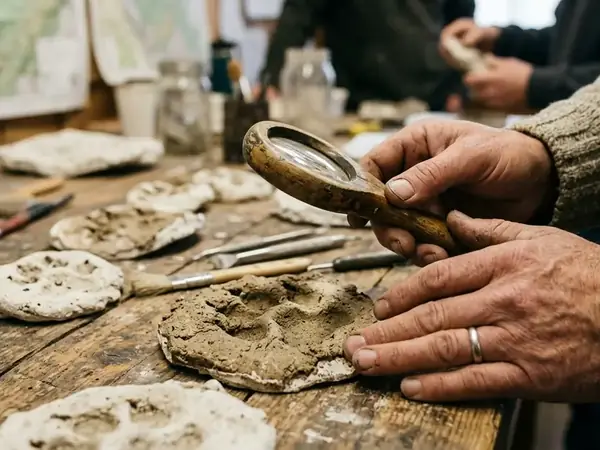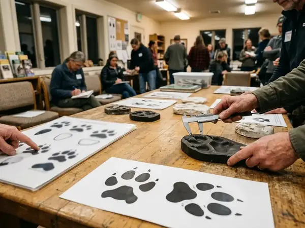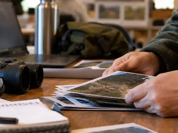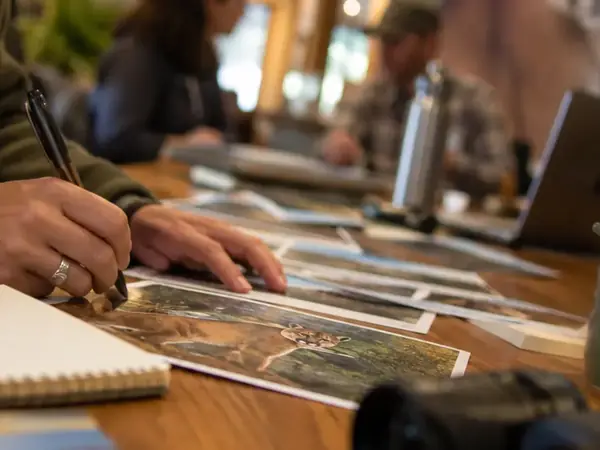

Kneeling beside a frozen Adirondack creek bed in late winter, I lay a metric scale next to a wind-scoured snow impression. Yesterday's gusts rounded the toe walls and blurred the leading edge of the heel pad. In crisp powder, distinguishing a wandering cougar from a big, splayed-foot bobcat comes down to stride, straddle, and pad geometry. Here in the crust, physical morphology fails us. Rather than walking away from an ambiguous print, we treat the depression as a biological sampling lead—photographing the impression with the scale, logging the overall trackway, and preparing to collect the snow itself for DNA analysis.
A Lone Print in the Adirondacks and the Cellular Trail Behind It
Every carnivore moving through an eastern hardwood forest leaves a microscopic wake. As an animal travels, it deposits genetic material through shed skin cells, saliva, urine, feces, and tissue associated with hair roots. Even when we find a single guard hair snagged on winter bark, the visible shaft alone may lack an attached root follicle rich in nuclear DNA, making the surrounding substrate just as valuable to collect.
Capturing this environmental DNA (eDNA) gives field biologists a practical way to screen vast, rugged eastern habitats where camera traps and visual sightings routinely fall short. A solitary cat can traverse an entire mountain range without stepping in front of a lens. By collecting the cellular remnants left behind on snow, soil, or water, we can identify specific drainages and ridgelines that deserve concentrated follow-up.
The snow-print-to-sequence chain relies on four linked steps: compressed paw-contact snow, a collection-matched blank, inhibitor-aware extraction, and independently checked cougar amplification. For that laboratory result to mean anything months later, the field notes must anchor it to reality. Before lifting a single gram of snow, record the sampled impression's GPS coordinates, the substrate condition, apparent wind or sun weathering, and its exact relationship to other tracks in the trail.
Collecting Genetic Material from Snow Tracks, Streams, and Soil
Where an animal travels dictates how we harvest its cellular trail. A snow track holds cells pressed directly into the crystalline matrix, whereas a forest stream carries suspended material washed off an animal's coat or tracks upstream. We keep snow, water, and soil sample types completely separate, matching the collection tool to the physical matrix.
Scraping Winter Trackways
When sampling a suspected pugmark in snow, timing matters. Direct sunlight, mid-day thawing, and wind disturbance rapidly degrade exposed genetic chains. After photographing the documented portion of the print, I use a sterile scoop to shave only the compressed contact surface and immediately associated snow, sliding it directly into a sterile, leakproof container.
Filtering Riparian Crossings
In warmer months or along open-water corridors, riparian pinch points offer another collection avenue. Following standard U.S. Geological Survey environmental DNA field protocols, we pump cold stream water through a microporous filter—such as a 0.45-micrometer nitrocellulose membrane, rather than a coarse mesh designed to sieve out whole organisms. Because waterborne material moves downstream, the field log must note upstream tributary junctions and our exact sampling position relative to suspected bank crossings, along with the total water volume filtered and any sediment clogging.
Swabbing Scrapes and Forest Litter
Away from water and snow, we target probable scent-marking scrapes, dry rock overhangs, and suspected feeding sites. Here we either swab the contacted surface or gather a small substrate sample into a sterile tube.
Eastern deciduous forests present a harsh chemical environment for shed cells. Damp leaf litter supports intense microbial and enzymatic activity that chews through free DNA, while humic compounds leached from decaying hardwood leaves frequently carry over into the lab extract and interfere with enzymatic amplification. Winter freezing slows many of these breakdown processes, which is why a cold, dry rock ledge or frozen soil scrape often yields cleaner material than damp summer humus. Whenever we sample forest floor material, we log substrate type, moisture level, and whether the spot represents an active scrape, a kill site, or ordinary background litter.
Guarding Against Field Contamination and Degraded Samples
Trace genetic sampling demands relentless discipline because the PCR reactions used in the lab are sensitive enough to amplify a single stray cell. Field collection must follow a strict clean-to-dirty workflow:
- Label all sterile sample containers before opening any gear near the track or stream.
- Put on a fresh pair of single-use nitrile gloves for every individual sample collected.
- Use single-use disposable scoops and forceps, or run metal tools through a validated bleach decontamination procedure followed by an appropriate rinse to destroy residual nucleic acids.
- Keep used tools and filled vials completely segregated from clean supply bags.
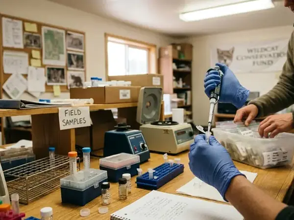
To verify that our field hygiene held up, we carry negative field blanks through the exact same physical steps as our environmental samples. A sterile water bottle merely uncapped to ambient air checks a very different exposure pathway than sterile water pumped through filtration tubing, a filter holder, or poured across a sampling scoop. Matching the blank to the specific collection procedure is the only way to demonstrate that the collector did not introduce foreign DNA.
DNA Analysis Field Hygiene: Leave domestic dogs at home and clean field boots between watersheds. A hiker's tread or a handler's gear can easily transfer canid biological debris or old scat traces straight into a pristine sampling site.
We also have to separate collector-mediated contamination from natural ecological transport. If a scavenging raven or coyote carries tissue fragments from a winter-killed deer across a watershed boundary, any cells dropped along the way are genuinely environmental DNA. However, that transport changes the spatial relationship between our collection point and the predator's actual location.
Finally, every field researcher must respect the biological limits of degraded substrates. Heat, ultraviolet radiation, and microbial enzymes cleave DNA strands into tiny fragments. Drawing on a review of DNA analysis records, a negative assay result indicates only that intact target DNA was absent from that specific spoonful of soil or liter of melted snow—never that the animal is absent from the surrounding forest.
Inside the Lab: Primers, Amplification, and Species Identification
Once the samples reach the genetics facility, technicians extract DNA from the nitrocellulose membrane, melted snow precipitate, or soil matrix using a chemistry tailored to strip out soil inhibitors like humic acid. Because environmental DNA arrives in the lab heavily fragmented, conservation geneticists target short regions of mitochondrial DNA (mtDNA). Every cell contains hundreds of mitochondria compared to a single nucleus, and short targets have a much higher chance of surviving intact across the span where primers bind.
To hunt for eastern cougar evidence, the lab runs Polymerase Chain Reaction (PCR) or quantitative PCR (qPCR) using candidate Puma concolor primers and, in probe-based qPCR setups, a matching fluorescent probe. Geneticists design these short nucleotide sequences by comparing reference genomes, then validate their specificity in the laboratory against closely related sympatric carnivores, especially bobcat (Lynx rufus) and domestic cat (Felis catus). The primers must latch exclusively onto cougar sequences while ignoring a bobcat track from the same ledge.
Quality Control and Assay Limits
Low-copy genetic work is prone to laboratory artifacts, so rigorous validation happens long before we interpret an exciting field sample. Every batch requires extraction blanks, no-template PCR controls, positive controls kept physically isolated from low-copy sample prep rooms, and an internal inhibition check to verify that forest tannins did not silence the reaction.
Sometimes primers interact with one another in the tube to form primer dimers—short, non-specific amplification artifacts that can mimic a weak positive signal. Analysts separate true cougar detections from artifacts by checking product size, examining melt-curve behavior, monitoring probe fluorescence, and sequencing the amplified product directly. Labs also run multiple independent replicate reactions from the same extract, recording the exact number of replicates tested alongside the number producing a qualifying signal against pre-established validation rules.
Even when sequence confirmation locks in a match, we have to remember what a mitochondrial assay actually measures. A species-level Puma concolor test identifies cougar DNA in the tube; determining whether that animal originated from a captive-release background, a dispersing western population, or identifying it as an individual requires additional nuclear genetic markers that are rarely recoverable from trace snow or water.
Why Genetic Hits Require Camera Traps and Physical Verification
A verified eDNA detection is the beginning of a field investigation, never the conclusion of one. At the scale of a sample vial, a positive hit proves that target DNA was present in the collected substrate, which does not establish a resident breeding population. A lone juvenile male dispersing hundreds of miles, predator scat carried in on a boot or coyote coat, or historical DNA preserved in cold, anoxic sediment can each separate a molecular detection from present-day residency.
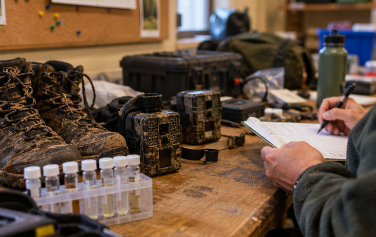
From Adirondack creek DNA to limestone-ledge cameras, the workflow is deliberate: use a molecular detection to place the next investigation, then seek repeated occupancy and reproduction evidence. When a lab reports a sequence-confirmed hit, we pull the original field notes and transport pathway records to design a geographically explicit follow-up grid:
- Stream detections: Because water carries cells downhill, we trace upstream tributaries and map likely bank crossings rather than strapping every camera to the trees right at the water-collection bottle site.
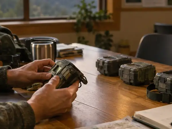
- Track and ledge detections: We concentrate trail camera arrays, permitted hair-snare stations, and scat-detection dog routes along the topological funnels—ridgeline saddles, rimrock ledges, and deer wintering yards, connected to the original trackway.
- Data linking: Every follow-up camera station and search transect is logged under the original eDNA sample identifier so that subsequent photographs, recovered scat, snag hairs, and lab genotypes can be evaluated as a single body of evidence.
Only by layering these methods can we answer the distinct ecological questions that matter for eastern carnivore recovery. Repeated photographs over multiple seasons demonstrate continued use of a territory; nuclear genotypes from fresh scat or hair snares distinguish individual animals; documented kittens confirm local reproduction.
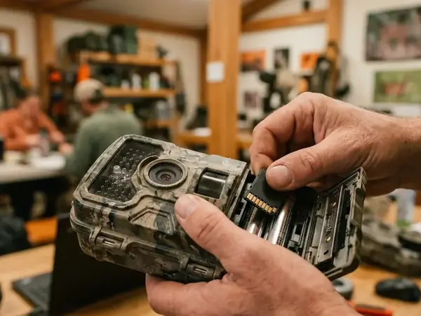
Late on a crisp November afternoon, I tighten the cap on a labeled vial of melted snow scraped from the shadow of a remote limestone ledge. After tucking the tube into an insulated pack and logging the GPS waypoint, I walk twenty paces across the hemlock bench. I cinch a trail camera strap around a straight trunk, aim the lens back toward the narrow rock shelf, and step out of the frame—leaving the glass eye to watch for the animal that left its cells in the frost.